Stages of Development During Beef Cattle Pregnancy
Bruce B. Carpenter & L.R. Sprott
Download the PDF | Email for Questions
Increasing Herd Productivity | Palpating to Determine Pregnancy | Developmental Stage | Palpation Technique | Determine Stage of Pregnancy | Caution | Other Factors | Systematic Determination | Recommendations | Other Methods
Determining pregnancy in cattle is an important management tool. The ability to determine pregnancy can allow you to make timely culling decisions and focus the resources of your operation on sound, reliable breeders. With experience, you can determine the fetal age which will allow you to predict expected calving dates and plan for the necessary labor at calving time. Pregnancy determination can also help you manage feeding to meet the high nutritional demands of gestation, calving, lactation and rebreeding more effectively.
Knowing expected calving dates can also be an advantage when marketing bred replacement heifers. Potential buyers often want to purchase females whose calving dates coincide with those of their present herd.
Increasing Herd Productivity
A cow-calf producer's economic returns depend largely on the percent calf crop and the weaning weight of the calves to be sold. You can compute your calf crop percentage by dividing the number of calves raised to weaning age (7 months) by the number of cows in your herd at the start of the breeding season.
Percent Calf Crop and Weaning Weight
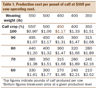
Table 1 shows the cost per pound of calf produced for various production levels with an operating cost of $550 per cow per year. To determine the required level of production for this example, take an arbitrary selling price of $1.31 per pound and locate the break-even point in Table 1.
Weanling calves weighing 450 pounds would require a 90 percent calf crop to break even. If only a 60 percent calf crop is produced, then the needed break-even would be 65 cents higher than the $1.31 market value. Calves weighing 500 pounds would break even at an 80 percent calf crop, and calves weighing 550 pounds would break even at just over a 70 percent calf crop.
Or, viewing it another way, lightweight calves averaging 350 pounds with only a 60 percent calf crop would need to sell for $2.52 per pound to break even.
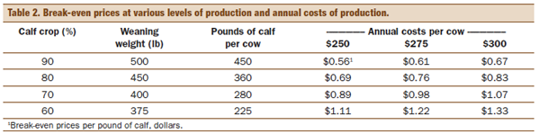
You can see the economic importance of calf crop and its interaction with weaning weight in Table 2. Clearly, break-even prices decrease as calf crop and weaning weight increase. This is true under any annual operating cost per cow.
Management Practices To Improve Production
The challenge for cow-calf producers is to use management techniques that stimulate production without drastically increasing operating costs. You can improve weaning weight through a number of methods, including:
- Internal parasite control.
- Using growth stimulants.
- Using sires with the genetic potential for increased growth.
- Providing adequate herd nutrition which also helps optimize reproduction.
Another effective and inexpensive way to improve reproduction is through annual pregnancy testing and culling of subfertile cows.
 In addition, culling open cows (and sometimes even bred cows) during an extended drought, may be necessary in order to balance stocking rate with declining forage supplies. However, there may be unique years when a drought during the breeding season is followed by rain later in the year. If forage conditions improve significantly, so may chances for re-breeding. Under such circumstances, "low risk" cows (e.g., middle-aged cows that are physically sound and proven breeders) are sometimes kept over until the next breeding season and given a second opportunity. This is also an acceptable option if replacement costs are higher than the cost of retaining the open cow. Remember that open heifers and aged cows are "high risk."
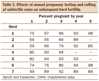
They should probably never be allowed a second breeding opportunity. Regardless of environmental considerations or replacement costs, any cow that is open more than once should be culled. If you follow this practice annually and your cows are under otherwise good management, pregnancy rates will increase. Table 3 shows that the increases in the pregnancy rate in Texas test herds were sustained at an acceptable level.
 Visual observation is also important in culling decisions. Some pregnant cows should be culled on the basis of age. Other determining factors are conditions of the udder, feet, legs and teeth that make them poor breeding stock. The decision to cull an open cow may also depend on her reproductive history.
Culling decisions may also depend on the animal's monetary value. You can retain highly valued registered females that fail to conceive until some or most of their initial cost is recovered. This strategy is prudent as long as these animals are free of abnormalities of the reproductive tract and have previously produced at an economically acceptable level.
Palpating to Determine Pregnancy
Pregnancy determination, or palpation, is made by inserting the arm into the rectum and feeling the reproductive tract for pregnancy indications.
Equipment
Little equipment is needed in palpation. The palpator should wear a protective plastic sleeve that covers the arm and hand up to the shoulder. The sleeve guards against disease and prevents irritation of the arm. Use an obstetrical lubricant or mineral oil to make entry into the rectum easier. Don't use soap or detergents as a lubricant, since both are irritants. Plastic sleeves may tear after several uses, reducing protection. If the sleeve tears, replace it before palpating the next animal.
The chute for holding the animal during palpation should allow her to stand in a normal position. It should have a front wall or gate and a bar just above the animal's hocks in the rear (Figure 1). This bar keeps the cow from kicking and protects the palpator. Include an entrance gate in the chute at the rear of the animal to allow the palpator to enter and exit easily. Provide a gate to swing across the crowding chute and fasten it in front of other animals coming behind the palpator. You may use squeeze chutes, but do not need to catch the cow's head for this procedure.
Palpation alone takes only a few seconds. The speed of pregnancy determination depends on three factors: management of the cows as they come through the chutes, the stage of pregnancy and the palpator's experience. Ideally, an experienced palpator can examine several hundred head of cattle in a normal working day. However, the process is much slower if the palpator has to help bring the cattle into the chute or climb over the chute wall to get behind the animal to palpate her.
To ensure the safety of the animals and the palpator, you must practice certain precautions:
- Restrain the animal so she cannot jump over the side of the chute or kick the palpator.
- Prevent other cattle from coming up behind the palpator as he or she attempts to determine pregnancy.
- Replace broken boards in the chute that could injure the animal's legs and check for exposed nails.
- Place cleats across the floor if it is slick to help stabilize the animal's footing
These precautions make it much more likely that the cow will remain calm and stand quietly during palpation. In addition, the process becomes more efficient and safe for the cow and the palpator.
Reproductive Tract Structures
Thorough knowledge of the structures of the female reproductive system is essential for successful palpation. Only the reproductive tract and associated organs will be discussed here, but you should be aware that endocrine glands located in other parts of the body, particularly the brain, are also involved in the sexual cycle. Figure 2 is a general diagram of the reproductive tract.
The vulva is the external portion of the reproductive tract and can be seen as two prominent lips. Its size may vary with the animal's age and between breeds. Brahman-influenced females usually have a larger vulva than do cattle of English and European breeds.
Figure 2 (right to left) shows the next portion of the tract: the vagina. It serves as a receptacle for semen during natural mating, is a thin-walled structure and is not easily felt during palpation.
The urinary bladder (not shown in Figure 2) is underneath the vagina. It may extend beyond the pelvic brim and slightly into the body cavity, particularly when it is full of urine. During urination, the bladder empties through a small opening (urethral orifice) on the floor of the vagina, eventually exiting the body through the vulva.
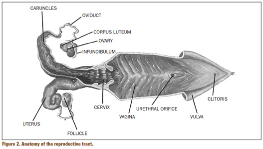
The cervix is a thick-walled structure attached to the vagina. It is comprised of connective tissue, which feels much like gristle. Because of the thickness and firm feel, the cervix is a good landmark for orientation while you are palpating. The internal walls of the cervix are folded and protrude toward the exterior of the reproductive tract. These folds are sometimes called cervical rings. The surface of these rings is lined with special mucus-secreting cells. This mucus is often seen smeared on the rumps or flowing out of the vulva of cows in estrus. During pregnancy, the mucus is much thicker than at estrus and plugs the cervix. This protects the developing embryo from foreign debris in the vagina. The cervix may also act as a sperm sieve, trapping some abnormal sperm cells and allowing normal sperm cells to travel into the uterus and oviducts.
The uterus is Y-shaped with a right and left horn. The horns share a connecting region known as the body. During artificial insemination, semen is deposited in the uterine body. The walls of the uterus are lined with special glands that secrete uterine milk, the substance that nourishes an early embryo. By approximately 16 to 18 days of gestation, the placental membranes are well developed and extend into both horns of the uterus. About 38 days into gestation, these membranes begin attaching to the uterine wall at special, raised areas known as caruncles. Located throughout the uterus, these are the exchange points for nutrients coming from the dam. The placental side of these attachment points are called cotyledons. The cotyledon with the caruncle is forms a combination known as a placentome, or button. In mid- and later gestation, these buttons become firm and are easy to detect when you are palpating the uterine surface.
The end of each uterine horn is attached to an oviduct, also known as a fallopian tube. The oviducts are small, tube-like structures. Because they are very small, they are difficult to feel. They transport sperm cells to the site of fertilization (the upper third of the oviduct) and an embryo back to the uterus if conception occurs. At the end of each oviduct is a thin, cup-like membrane that is difficult to feel: the infundibulum. It catches the egg, or ovum, as it is expelled from an ovarian follicle during ovulation, and transports the egg into the oviduct for eventual fertilization. That is why each ovary is located close to the infundibulum.
The entire reproductive tract is attached to a thin suspensory membrane known as the broad ligament. This elastic-like ligament will stretch and move within the pelvic and body cavities to allow the reproductive tract to move. This movement is necessary because of the weight of the fetus and the crowding of the tract by other internal organs. The broad ligament acts as a cradle for the tract and is attached to the upper pelvic and body cavities. It also contains arteries and veins that supply the tract with blood to nourish the tissues.
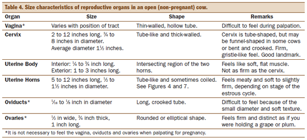
Figure 3 shows an interior view of the reproductive tract and broad ligaments of an open (non-pregnant) cow. Table 4 gives a general size description of the various parts of the tract in an open cow. The complete tract with all its parts varies in size and feel. This depends on the stage of the estrous cycle and on the breed, size, and reproductive history of the animal. Generally, the size of the entire open tract is 12 to 18 inches long. In young heifers that have just reached puberty, the tract may be only 8 inches long. The tract of mature cows that have had several calves may extend to 24 inches.
 Changes Associated with Pregnancy
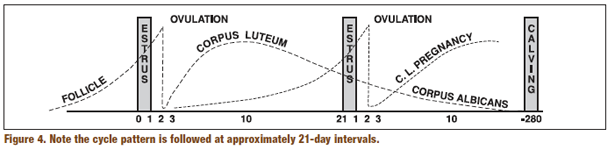
The sexual cycle of a normal cow is characterized by estrus, or heat, periods that occur at 21-day intervals. Figure 4 diagrams the estrous cycle and some of its activities.
At estrus, the cow is influenced by estrogen being produced by follicles on the ovaries. This hormone causes her to display estrus. Within 24 hours after the initial stages of estrus, one of the follicles ruptures and releases a single ovum, or egg. This is ovulation. The egg moves into the infundibulum and eventually down into the oviduct. The cavity on the ovary left by the ruptured follicle develops into a new structure known as a corpus luteum (Figure 2). The corpus luteum produces progesterone, the hormone responsible for maintaining pregnancy. If conception does not occur with this ovulation, the uterus releases a hormone called prostaglandin. It regresses, or destroys, the corpus luteum.
Regression is complete by approximately 16 to 17 days into the cycle. Meanwhile, follicles continue to grow on the ovary. Since the corpus luteum has regressed and is no longer producing progesterone, a new ovulatory follicle is recruited. Within 4 to 5 days, the cow returns to estrus. The process of development and regression of a normal corpus luteum causes a cow to have her characteristic 21-day sexual cycle.
If the cow is mated during estrus, the sperm cells will travel from the site of deposition (the vagina) to the site of fertilization (upper third of the oviduct) within minutes. While there, the sperm cells undergo a 6- to 8-hour maturation period called capacitation. Only then can the sperm cells fertilize an egg. Consequently, when the egg arrives at the fertilization site, the sperm cells are already there and probably have undergone capacitation. The chances of fertilization and pregnancy average about 50 to 70 percent.
When fertilization occurs, the cow's physiological cycle begins a dramatic change. This leads to the development of a full-term fetus. About 8 days after fertilization, the embryo is transported to the uterus. At 16 to 17 days after fertilization, the embryo and its placental membranes begin to release a hormonal signal that prevents the usual release of prostaglandin from the uterus. As a result, the corpus luteum does not regress and continues to release progesterone to maintain the pregnancy. The embryo houses itself in the uterine horn nearest the ovary that produced the ovulating follicle. Therefore, an embryo found in the right horn came from an egg produced by the right ovary, and vice versa. For this reason, particularly in cows that are open or may be in the early period of gestation, you must palpate both uterine horns.
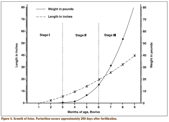
The major changes in the structure of the reproductive tract after conception occur mainly in the uterus. Its shape, size, texture or feel, and location will change. Also, the embryo will grow, and its growth is directly responsible for changes noted in the uterus. Figure 5 shows embryo or fetal growth by month and stage of gestation.
Developmental Stage
There are three main periods of development in a young calf's life. The period of the ovum is that time from fertilization until the egg has divided enough times to take on a particular form. This occurs on approximately the thirtieth day when there is an enfolding of the layers of the developing egg. At this stage the newly developing animal is called an embryo. The period of the embryo lasts until the fetal membranes begin to attach to the lining of the uterus which takes approximately 38 days. During the embryonic stage, various organs and systems are laid down. These include the respiratory, nervous, digestive, circulatory and reproductive systems. As the embryo develops, it floats freely in uterine milk in the uterine cavity.
The fetus period begins when the embryo is approximately 38 days old and ends when the newborn is expelled at parturition, or birth (Figure 5). During the fetus stage, continued attachment takes place at the numerous caruncles lining the uterus. These attachments provide transfer of nutrients and waste materials for the developing fetus. Birth occurs approximately 280 days after fertilization.
Palpation Technique
You may use either hand in palpation. With one hand, you may grasp the cow's tail as a handle while you palpate with the other. The hand used for palpation should be well lubricated and shaped into a wedge by bringing the fingers together as closely as possible.
Rectum
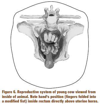
Push through the anus into the rectum with one continuous thrust. As your hand enters the rectum, fold your fingers into a modified fist by tucking under the fingertips (Figure 6).
In this position, your hand will push the fecal material aside and straighten the rectum. Folds in the rectum do not straighten as easily if the fingers are pointed. The modified fist also eliminates the risk of puncturing the rectal wall with sharper, pointed fingers. However, puncturing is rare, as the rectum is thick-walled and resistant. Cleaning the cow's rectum of fecal material usually is unnecessary. However, in early stages of learning, cleaning the rectum will help you feel other structures more easily.
 Rectal straining may result from the entry of your hand. This is a reflex response by the cow. Straining can be alleviated by simply moving your hand back and forth in a gentle massaging motion. If straining reoccurs, massage the rectal wall again.
Usually, the longer the examination, the more rectal straining you will encounter. Do not be upset by a small amount of bleeding, as this occurs occasionally and is not necessarily a sign of damage to the rectum. An indication of rectal damage is a sandpaper or gritty feeling, which means that the mucosa lining of the rectum has been rubbed off during palpation. If this occurs, it is best to stop palpating immediately.
Feeling through the rectal wall is similar to feeling through one or two layers of thin rubber. Most cattle are cooperative, so you should be able to detect and pick up the reproductive organs easily.
Experienced palpators usually learn to follow a consistent routine. When first entering the rectum, thrust the arm beyond the elbow and deep into the abdominal cavity. Feel in a downward direction toward the udder. Since late-term fetuses will be located here, initial deep entry allows palpators to determine pregnancy quickly and reduces time spent in the cow. If a fetus or other indications of pregnancy are not found in the abdominal cavity, palpators should move back toward the pelvic cavity. Most open tracts and early pregnancies will be located here. While feeling in or near the pelvic cavity, locate the pelvic brim. This is a good landmark for orientation. Most importantly, find the cervix and move forward to the uterus to determine pregnancy. Following this simple routine will help reduce palpating errors.
 Paunch
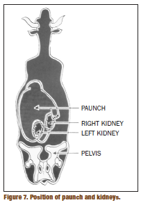
Upon entry into the rectum and just past the pelvic brim, palpators may encounter the paunch, or rumen. It is the first and largest of the cow's four stomachs (Figure 7). It has a dorsal (upper) and ventral (lower) sac. The dorsal sac is forward and to the left of the pelvic brim. It may feel like the end of a football and be mushy or gritty. This protruding end of the dorsal sac may even extend into the pelvic cavity. In such cases you might mistake it for an enlarged uterus or late-term fetus. By mashing the paunch, you will notice an indentation which gradually smooths back to its original shape. This indicates that the paunch is full of feed. The paunch does not have the watery feel like a pregnant uterus.
The ventral sac of the paunch is deep into the body cavity and located toward the udder region. This sac lies either to the right or to the left side of the body, depending on your gut fill. It has the same mushy or gritty feel as the dorsal sac and will indent if mashed with the hand.
 Reproductive Tract
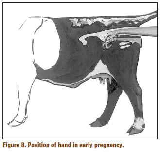
The open reproductive tract normally lies on the pelvic floor or against either pelvic wall. The horns of this tract are usually coiled on their front edge and may hang slightly into the abdominal cavity (Figure 8) in older cows. Two characteristics confirm the absence of a pregnancy: (1) when palpated, both uterine horns in the open cow will feel thick-walled and have a meaty texture; and (2) fluid will not be in the horns. At this stage you can hold the entire uterus in your hand and palpate it from underneath or from the side. However, when learning to palpate, it may be easier for you to feel the top surface (Figures 8 and 9).
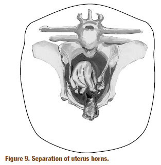
Tone, or firmness, of the open horns varies with the estrous cycle. Shortly before and after estrus (under estrogen influence), the uterus will feel turgid or firm. During the period of the corpus luteum (progesterone influence), the uterine horns will feel flaccid or soft. Slight pressure with the middle finger (Figure 9) will separate the horns, allowing you to palpate each horn. Both horns must be felt, since a pregnancy may occur in either. Inexperienced palpators sometimes have difficulty grasping the uterine horns. To overcome this problem, move the uterus down against the pelvic floor or to either pelvic wall. Flatten your hand and apply gentle pressure against the uterus to separate the horns.
 The ovaries are in the broad ligament toward the end of the uterine horns or at their sides. However, it is not necessary to palpate the ovaries when determining pregnancy. The most important step is to feel the uterus for its texture and content.
Determine Stage of Pregnancy
Stage I: 30- to 35-day pregnancy
Because embryos at this early stage are delicate, beginning palpators should not try to feel them. An experienced palpator, however, can detect pregnancy as early as 30 days after breeding. Palpation which at this early stage should be accompanied by good breeding herd records. These records help the palpator know the approximate breeding date of the animal.
In the early stage of pregnancy, the uterus is filled with a small amount of fluid and will feel slightly thin-walled. One horn is enlarged a little more than the other. At this stage you can determine the presence of the embryonic vesicle by carefully running the horn between your fingers in a milking action. You can feel the vesicle slide through your fingers.
At this stage the embryo is only about 1⁄2 inch long. The vesicle surrounding it is about 3⁄4 inch in diameter and filled with fluid—like a balloon filled tightly with water. However, the borders of this vesicle are indistinct. What you actually feel is something slightly smaller than a marble as it slides through your fingers. The uterus, in much the same location as a non-pregnant uterus, has not been displaced because of size or weight at this time. The outer embryonic vesicle, which occupies both horns, is rather thin with little fluid. It may be 18 to 24 inches long. By pinching the horn of the uterus carefully, you can feel the membranes of this vesicle as they slip between your fingers.
Stage I: 45-day pregnancy
Most palpators prefer that bulls be separated from cows at least 45 days before pregnancy determination. At 45 days, the horn of the uterus containing the fetus is somewhat enlarged and thinner-walled compared to the other. The fetus at this stage is about 1 inch long. The vesicle around it is egg-shaped and measures about 1 to 11⁄2 inches long. You can feel the outer membrane, which contains fluid, through the uterine wall. The attachment of the membranes to the uterus has just taken place at about 38 to 40 days. Therefore, avoid moving the fetus about in the uterus. The caruncles on the uterus join the cotyledons, on the fetal membranes for nutrient exchange. These two structures form the placentome. Buttons cannot be felt by palpation until later in pregnancy.
Slipping of the fetal membranes is a valuable aid to early pregnancy determination. Although the membranes can be slipped at any stage of gestation, it is easiest to perform and of the most value between 40 to 90 days of pregnancy. The procedure involves picking up and gently pinching together the walls of either uterine horn. The palpator feels the fetal membranes as they slip between the thumb and fingers. Be gentle when using this technique, since the embryo and membranes are rather delicate in pregnancies under 45 days.
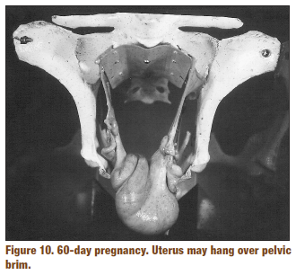
Stage I: 60-day pregnancy
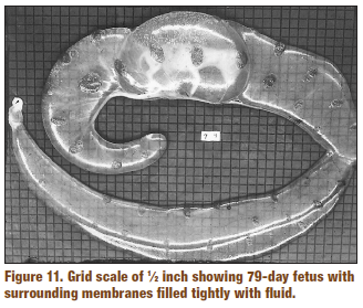
The uterus, which will have enlarged considerably by this time, is filled with fluid and increased growth of the fetus (Figure 10). The fetus now is about 21⁄2 inches long and may have displaced itself into the abdominal cavity, indicating that the uterus has stretched. The cervix may be pulled over the pelvic brim; but the cervix, body and horns of the uterus are within reach. In larger animals, this is a difficult stage for pregnancy determination. This is due to displacement and the distance from the anus to the developing fetus.
The uterine walls have thinned considerably. The best method of feeling the fetus is to bobble it with your hand so that, if you gently tap the uterus, the fetus swings like a pendulum and hits against the wall of the uterus and vesicle. The cervix remains on top of the pelvic cradle, and the uterine horns move toward—and possibly beyond—the brim.
 Stage I: 90-day pregnancy
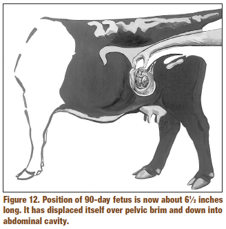
The fetus now is about 6 inches long and may have displaced itself into the abdominal cavity, indicating that the uterus has stretched (Figure 12). The cervix may be pulled over the pelvic brim; but the cervix, body and horns of the uterus are within reach. In larger animals, this is a difficult stage for pregnancy determination. This is due to displacement and the distance from the anus to the developing fetus.
You may have to consider factors other than the presence of the fetus at this stage. Consider displacement of the uterus as a possible indication of pregnancy. Another indication of pregnancy is enlargement of the uterine arteries with their characteristic pulsation. These arteries (one for each uterine horn) are in the forward fold of the broad ligament (Figure 13), which supports the uterus. In a three-month pregnancy, the artery supplying blood to the pregnant uterine horn is about 1⁄8 to 3⁄16 inch in diameter. The artery feeding the non-pregnant horn is only half that size. When you grasp the artery, you can easily feel the pulse of the heartbeat as blood is carried into the uterus to nourish the developing fetus. Do not confuse the uterine artery with the femoral artery lying on the inside of the thigh which supplies the hind legs. The femoral artery is in the muscle, but may be palpated. Remember that the uterine artery is in the broad ligament and can be moved 4 to 6 inches, while the femoral cannot.
 If you are unable to reach the fetus, the best indication of pregnancy is the presence of buttons, which are now large enough to palpate. In a 3-month pregnancy, they are flattened and egg-shaped and measure 3⁄4 inch to 1 inch across. At this stage they are still rather soft to the touch, but they are firmer than the thin-walled uterus. The membranes still are filled tightly with fluid.
Stage II: 120 days until 6 months
At approximately 100 days, buttons become more apparent (1 to 11⁄2 inches) and can usually be palpated. At this stage the fetus is still displaced similarly to the 90-day fetus. However, it has grown to approximately 10 to 12 inches long, with the head almost the size of a lemon. Often, the palpator can detect the head of the developing fetus before any other body part.
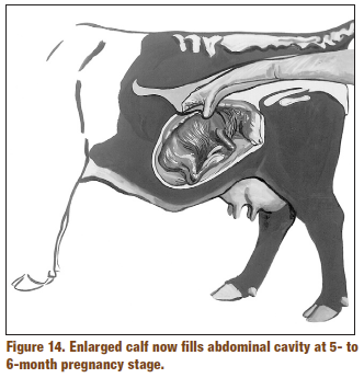
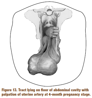 As the fetus enlarges and grows (now 4 to 6 months), its sheer weight displaces the entire reproductive tract deeper into the body cavity. This requires palpation deep in the body cavity (Figure 14). All other characteristics have also changed. The buttons are more noticeable, since they have developed to about 2 to 21⁄2 inches in length and have a much firmer feel. The pulsating uterine artery may be palpated (Figure 13) and is now about the size of your little finger.
Stage III: 7 months until 9 months
At 6 to 7 months, the fetus may or may not be deep in the body cavity (Figure 14). Remember to reach deep and toward the stomach floor.
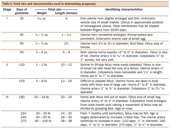
 By 7 to 9 months, the fetus has grown to where it is often easy to palpate without reaching deeply. You can often feel the large bony structures (e.g., head, legs and back). You may feel fetal movement. Occasionally in large bodied cows, the fetus may still be completely out of reach. To help confirm pregnancy, look for well developed buttons or weight on the displaced cervix. You can also check the uterine arteries, which are now about the size of your thumb. The main change until parturition will be in size, as the fetus grows rapidly and uses more of the abdominal cavity. Table 5 summarizes outstanding identifying characteristics.
Caution
One word of caution is necessary to help avoid errors in determining pregnancy. The position of the reproductive tract, in and of itself, is not a reliable way to determine pregnancy. Many 90-day (and shorter) pregnancies may be located entirely in the pelvic cavity and not be displaced beyond the pelvic brim. On the other hand, pregnancies beyond 90 days are big enough that they most often are displaced beyond the pelvic brim. Conversely, some open tracts are not always located entirely within the pelvic cavity. In large-frame, fat cows, the open uteri may fall beyond the pelvic brim. In all cases, the tract must be adequately traced to correctly determine pregnancy status. Simply stated, the location of the tract should be used as a roadmap to lead the hand to the uterus, whether it is displaced beyond the pelvic brim or not.
Other Factors
The paunch. Remember that the paunch (Figure 6) is often encountered when entering the rectum and feeling beyond the pelvic brim and toward the left or very low in the body cavity. The shape of the dorsal and ventral sacs may be mistaken for the head or rear quarters of a calf. The difference can be determined by mashing on these large objects. The paunch will indent when mashed, while a well developed calf may move away from the pressure of your touch. Also at these late stages of pregnancy, you can easily distinguish fetal features (e.g., ribs, hooves and ears) when you touch them.
Kidneys. The kidneys (Figure 6) are suspended directly under the spinal column at about a 30-degree downward angle. Because the left kidney in cattle is more toward the rear of the animal than the right one, it is often touched during palpation. The left kidney is elliptically shaped and is sometimes mistaken for a calf's nose. Practice helps you to distinguish the difference, but inexperienced palpators can avoid the left kidney by feeling at a steeper angle into the abdominal cavity. It is usually at this steep angle that large fetuses are located.
Buttons. Buttons may be mistaken for ovaries or vice versa. Buttons do not have the solid feel of an ovary but are rather soft. The best comparison is that they feel like dried apricots soaked in water. The ovaries are more rounded and egg-shaped with a firm feel. Only two are present.
Pyometra. In this condition, the uterus is filled with white blood cells attempting to clear up disease organisms. The uterus may be fluid to the touch or may be somewhat solidified, feeling rather plastic. This stage may be confused with early pregnancy stages if the uterus is in a fluid condition and only partly filled. In the latter stages of pyometra, the uterus becomes rather firm.
Large uteri. In older cows that have had many calves, the uterus may not return to its normal size, as it will in a younger cow. The enlarged uterus may be displaced over the brim of the pelvis as in a 3 to 4 month pregnancy. In the open cow, careful manipulation of the uterus will allow you to determine that no fluid and no developing buttons are present. Relaxation of the broad ligament tends to cause a similar condition.
Bladder. A full urinary bladder may be interpreted as a pregnancy in the 60- to 75-day stages. The full bladder feels similar to the uterus filled with fluid. Careful tracing should allow you to determine if the structure is the bladder, where there is only one body. This will also help you determine if the structure is a pregnant horn of the uterus, where both horns can be palpated and traced back to the cervix.
Enlarged cervix. Some Brahman and Brahman crossbred cattle have an enlarged cervix that is firm and feels like a developing fetus in the latter stages. You can distinguish between the two by tracing the reproductive tract.
Breed differences. Because of their large size, certain Brahman crossbreeds, Santa Gertrudis, Charolais, Holstein and Brown Swiss cattle are more difficult to palpate in certain stages of pregnancy than the smaller European breeds. In the 3-to-4month stages, the uterus may have dropped so deeply into the body cavity that it is almost impossible to palpate. In these cases, pass your hand under the cervix and lift the uterus to feel the fetus itself. By lifting the uterus and quickly moving your hand down into the body cavity, you can locate the fetus by gently bobbing the fluid and the fetus through the wall of the uterus.
Brahman and Charolais breeds appear to have more tissue inside than smaller breeds. More folds of the omentum seem to cover the intestines, making it slightly more difficult to pick up the uterus. Charolais cattle seem to have less flexibility in the rectum. It is commonly harder to feel deep in the body cavity in these cattle, and lateral movement is somewhat restricted. In Holstein cows, the anal sphincter may be tight. This limits the deep entry into the body cavity necessary to determine the stage of pregnancy. In these cases, proficiency at mid-uterine artery palpation may be necessary (Figure 13).
The uteri of Brahman or Brahman-influenced heifers vary considerably. It is not uncommon to find 1,000-pound heifers with uteri measuring only 4 to 6 inches in length compared to a normal uterus, which would be 10 to 12 inches.
Highly finished cattle may be filled with fat which interferes with movement and feel. These cattle may be difficult to palpate. If you are uncertain, repalpate at a later date.
Systematic Determination
Once you understand the variations in location and size of the reproductive organs, your ability to determine pregnancy accurately depends upon a careful and logical check of the various reproduc tive and fetal structures. Using a systematic approach to checking each cow will ensure that you determine critical changes and variations in the location, size and feel of the reproductive organs. A systematic approach not only helps assure accuracy, but saves the palpator a lot of work. Figure 15 shows a systematic approach to determining pregnancy. While the system can be modified, it has proven to be a very functional approach for beginning and experienced palpators.
Figure 16 provides an outline of the important and critical factors that must be carefully checked and considered in determining the stage of pregnancy. If your pregnancy determination is to be accurate, you must consider all factors. The outline is basically an arbitrary division of pregnancy based upon the location of their reproductive organs and/or fetus. It is included to summarize the more important factors that indicate whether the cow is open or pregnant. Beginners should study carefully the outline and be familiar with the changes and variations that occur in each stage of pregnancy.
Even after following a systematic approach to palpation, some beginners will be unable to confirm pregnancy status on some cows. If this happens to you, simply wait 30 days and check these few again. If they are pregnant, their uteri will have grown which makes it easier to confirm their status. Never speculate on their status, as incorrect guesses can be costly.
Occasionally, cattle producers want an anticipated calving date (or known date of conception) from pregnancy records. The best way to determine it is by palpating in the early stages of pregnancy (90 to 100 days or less). These pregnancies are usually within the palpator's reach and are small enough to be sized. This allows a close estimate of anticipated calving date. In late pregnancies (120 days or more), fetal size can vary greatly among cows that actually conceived on or about the same day. Consequently, determining anticipated calving date (or day of conception) from records taken in late pregnancy is difficult and often inaccurate.
Recommendations
Practice! Experience is the key to palpation. In many instances, the ranch manager should not be the one to palpate but should supervise the operation and critically observe the cows. Unhealthy, unsound and undesirable types should be eliminated, as should open cows.
Shorten the calving interval by reducing the time during the breeding season when the bulls are with the cows. Cows that settle first are most adapted to reproduction. Wait about 90 days after the bulls are removed before you palpate. Most cows should conceive at the beginning of the season, and only a few will be short-term pregnancies. Cull as critically as is feasible for your operation. If every open, unsound cow can be removed, cull immediately. Remember, palpation is an art and a skill. It pays dividends to the person who uses it wisely.
Other Methods
Ultrasound
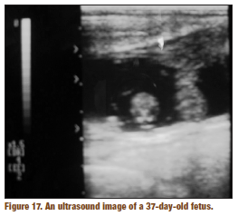
Trans-rectal ultrasonography has many uses for reproductive diagnostics in cattle, including pregnancy determination. However, using it as a method for routine pregnancy determination on animals in late stage I or in stages II or III of gestation is often not cost-effective. This is due to the high cost of ultra sound equipment and/or ultrasound service. Still, there are situations where ultrasound might be the best way to examine the uterus and/or conceptus. These might include early pregnancy determination, fetal sexing, or determining the number and/or viability of the conceptuses.
When performing a trans-rectal ultrasound examination, a trained person places a transducer probe in the rectum of the cow. Using the same hand to manipulate the probe and the uterus, the transducer sends and receives sound waves that have been directed along the uterine horns. Since tissues and/or fluid will reflect or absorb sound waves differently, those sound waves are transduced into an electronic image. This is then viewed on a special monitor. Skill is needed to maniuplate the probe and to interpret the image produced on the monitor (Figure 17). Pregnancy may be detected as early as 26-28 days, but examinations will be more accurate if they are preformed after 30 days.
Blood Test for Pregnancy Specific Protein B
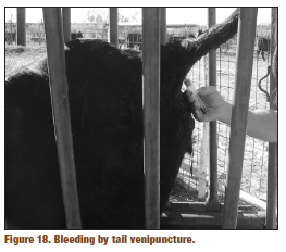
A new method to determine pregnancy became commercially available in 2004. Called the BioPRYN® test, it utilizes a laboratory procedure to test tail- or jugular-bled animals for pregnancy (Figure 18). It is essentially a "yes/no" test and is highly accurate: more than 99 percent on pregnant cows and about 95 percent on open cows. Blood collections are shipped by commercial carrier to a laboratory. Results are available by fax or e-mail within 27 hours after arrival at the lab. Cost of the test is $2.25 per sample plus purchase of blood collection equipment and shipping.
 BioPRYN® stands for pregnant ruminant yes/no, as it has application in many species of domestic and wild ruminant animals. The test uses an enzyme-linked immunosorbent assay (ELISA) that detects the presence of a protein known as pregnancy specific protein B (PSPB) in the blood of the mother. PSPB is produced by cells in the embryonic trophoblast or fetal placenta where it enters the into the mother's blood from the uterine caruncles. PSPB can be accurately detected as early as 30 days post-conception. (Cows and heifers, for example, can be tested as early as 30 days after breeding.) However, there is a caveat. Cows that have calved will have residual PSPB in their blood. Therefore, to allow for clearance time, it is recommended not to test lactating cows until at least 90 days after calving. When individual calving and/or AI records are available (e.g., dairy), individual lactating cows, could be tested as early as 90 days post-calving, assuming that breeding records also showed at least 30 or more days since breeding. With cows that are managed by pasture or herd (e.g., beef cows), the earliest that testing could be done would be 90 days after the end of the calving season. For example, a herd with a 90-day calving season would have a 90-day breeding season following that. Therefore, the earliest that testing could be done would be 30 days after the end of the breeding season. Testing can also be done anytime up until the end gestation.
 Cows are reported as open or pregnant based on the optical density of blood samples as measured by calibrated laboratory equipment. Although BioPRYN® is a "yes/no" test, some estimation of length of gestation can be made based on the relative magnitude of optical densities between samples. That is, animals in late gestation will have a relatively higher optical density score compared to animals that are in early gestation. Sometimes a cow will be reported as "open-repeat" or "pregnant-repeat." This indicates that the age of the embryo may be just less than 30 days or that it has died leaving some residual PSPB. It is recommended to bleed and test these animals again after a 5-day or more waiting period to allow for either embryo growth or clearance of residual early embryonic PSPB.
 When comparing BioPRYN® to rectal palpation or ultrasound, note that the results of the BioPRYN® test are not immediately available, so any keep or cull decisions would be postponed until receipt of the lab report. In addition, cattle must be individually identified to allow for culling or other management once the results of the BioPRYN® test are known. With the other two methods, neither individual ID nor the need to re-work cattle is required. Also, a skilled person can determine the stage of pregnancy (e.g., I, II or III) with either rectal palpation or ultrasound. This type of information may be useful for culling and marketing decisions, or in determining if conception may have occurred (unintentionally) after the end of a breeding season.
 Specialized uses for the BioPRYN® test are that it can be used in artificial insemination programs to determine conception to estrous synchronized AI versus clean-up bulls. To do this, clean-up bulls are not placed with the herd until 16 days after the last AI breeding. Blood testing at 30 to 35 days after AI will detect AI pregnancies but not early pregnancies to bulls. All cows or heifers classified as "open" with the first test, are re-tested 30 or more days after the end of the bull breeding season. For more information, contact BioTracking LLC (www.biotracking.com).
Return to Top
chenaultcritheing.blogspot.com
Source: https://beefskillathon.tamu.edu/determine-pregnancy/
0 Response to "Stages of Development During Beef Cattle Pregnancy"
Postar um comentário